Focused Ion Beam

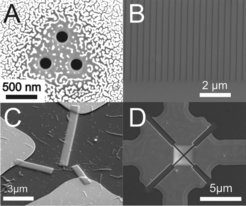
A versatile instrument to prepare and analyse nanostructures is the focused ion beam (FIB). In a FIB, a beam of ions is focused onto the surface and raster scanned. This allows the removal of material by ion sputtering (“milling”) with a resolution of ≈ 10 nm. The FEI Nova 600 Nanolab FIB instrument at the MPIP is a so-called dual-beam instrument that combines a focused ion beam with an SEM that allows simultaneous imaging of the sample. This combination is especially important for the preparation of delicate samples, where sample alignment is done under SEM control without the need of ion beam imaging. Our system is equipped with 3 gas injection systems that can be used for ion beam induced material deposition of Pt, Au or SiO2 layers. Further options include an EDX detector for elemental analysis, a nanomanipulator for the liftout of TEM-lamella (see below) and a cryo-system for investigations on “wet” samples as cells or hydrogels at temperatures down to -140°C. The main applications of the FIB at the MPIP are nanostructuring of surfaces, cross sections of samples, preparation, TEM sample preparation and cryo-SEM. The main application of FIB at the MPIP is the nanostructuring of surfaces. Fig. 1 shows examples nanostructures prepared by FIB milling.

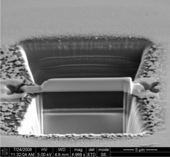
Where FIB opens completely new possibilities is the preparation of TEM lamellae, i.e. the site directed preparation of ~50 nm thin slices out of bulk samples that are hardly or not at all accessible by other means. Fig. 2 shows the first step in the preparation of a TEM lamella from a pixel of an OLED display. The lamella that contains the region of interest is already visible but has still to be cut out and lifted out with the nanomanipulator.
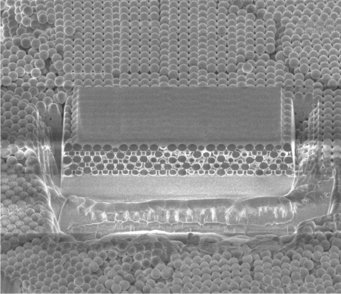
Using the so-called “slice and view” protocol, a 3D tomography of samples is possible. In this mode, slices of typically 10-100 nm thickness are removed by ion beam sputtering from a small block of the sample. After each slice, a SEM image of the newly exposed sample face is taken (Fig. 3). From such a set of slice images, a 3D reconstruction of the sample can be obtained by image analysis.


